User:Cortez713/sandbox
Article evaluation of Dorsal nerve cord[edit]
Week 2[edit]
Dorsal nerve cord:
- missing the evolutionary function and how the synapomorphy arose/developed
- needs to provide citations for formation and modification of dorsal nerve cord
- needs to provide citations for dorsal v. ventral, bipedal v. quadrupedal, the subphylum it belongs to, and the other 4 synapomorphies that Chordates possess
- due to lack of citations provided, the article would fall under the utilization of plagiarism
- why is Ectoderm capitalized? Did the wiki author mean to link it?
- in class, Dr. Schutz said we need to be concise and precise when explaining our answers but the article's sentences are drawn out and redundant
- not sure if discussing bipedalism and quadrupedalism and the difference between dorsal and ventral is entirely relevant
- lacking (cited) images that could be utilized to help the reader locate the dorsal nerve cord as well as help visualize the morphology
- It has not received any ratings of quality or of importance for WikiProject Biology, but for WikiProject Animal Anatomy, it has been rated Stub-Class and of low importance
Overview of structure:
- Overall, the article contains a neutral viewpoint, but the article committed plagiarism due to not citing and including references after the information used. The article lacked detail and further explanation of the topic as well. It was noted on top of the article's page that it did not cite any sources, but was not commented on in the Talk page. Sentences could be taken out due to relevance and be rearranged for clarity.
Week 3: Add/edit Dorsal nerve cord article[edit]
Added a citation to the Dorsal nerve cord article to the second paragraph that states: "a dorsal nerve cord is mainly found in subphylum Vertebrata. Chordates also usually have a notochord, a post-anal tail, an endostyle, and pharyngeal slits." As well as the third paragraph of: "dorsal means the "back" side, as opposed to ventral which is the "belly" side of an organism" and "in organisms which walk on four limbs the dorsal surface is the top (back) and the ventral surface is the bottom (belly). [1]
A citation to help support this would be: "a dorsal nerve cord is mainly found in subphylum Vertebrata. Chordates also usually have a notochord, a post-anal tail, an endostyle, and pharyngeal slits." [2] In addition, citatios would be added to: "dorsal means the "back" side, as opposed to ventral which is the "belly" side of an organism"[2] and "in organisms which walk on four limbs the dorsal surface is the top (back) and the ventral surface is the bottom (belly)."[1][3]
References[edit]
- ^ a b "Dorsal nerve cord". Wikipedia. 2018-01-18.
- ^ a b Kardong, Kenneth V. (2015). Vertebrates: Comparative Anatomy, Function, Evolution. New York: McGraw-Hill Education. ISBN 978-0-07-802302-6.
- ^ Sirois, Margi (2017). Elsevier's Veterinary Assisting Textbook. St. Louis: Elsevier, Inc. ISBN 9780323359221.
Week 4: Dissection organism assignment[edit]
- Stingray: It'd be interesting to learn how the venom works in concordance with the barbed stinger for self-defense. Also I'm curious if it's true that removing the barbed stingers on the rays are painless and how zoo's manage the venom glands when they allow the public to interact with them. Fish scale#Placoid scales
- Bat: It'd be interesting to study how it's morphology of its larynx is able to aid in producing echolocation and other vocalizations and see how closely related the structure of the throat is to humans and their ability to produce vocalizations. Vampire bat
- Turtle: I would like to understand how the turtle's body is constructed to allow its head and legs to retract into its shell to protect itself. Leatherback sea turtle
Week 5: Intro to dissection group (draft)[edit]
All contributions will be going towards User:Bucl003/sandbox.
I would contribute to the Rabbit page by adding a citation and more in depth information on rabbit ears in the morphology section that would discuss the outer and middle ear and how it aids the rabbit in having a more active lifestyle since the ears are utilized for sensory information.[1][2] Also, I would include information on the morphological and functional purpose of their eyes which are adapted for low light due to being active at dawn and dusk as well as the mouth that has adapted a certain structure to be able to eat short grass.[2]
Utilized links:
https://rabbit.org/rabbit-ears-a-structural-look-2
References[edit]
- ^ Rickel, Jana (Summer 2005). "Rabbit Ears: A Structural Look". House Rabbit Journal. 4 (11).
- ^ a b Aspinall, Victoria; Cappello, Melanie (2015). Introduction to Veterinary Anatomy and Physiology Textbook. China: Elsevier. ISBN 9780702057359.
Week 6: Rabbit article draft - pinna/outer ear[edit]
The Auricle (anatomy), also known as the pinna is a rabbit's outer ear as depicted by Fig. 1 to add an example to User:Bucl003/sandbox. [1]
The bones (humerus, radius-ulna, and paws) involved in the hindlimbs can be added to [[User:ReallyCaffeinated]] example by utilizing Fig. 29.18 or what would be our Fig. 2.[2]
The rabbit's body surface is mainly taken up by the pinnae. Their ears, utilized for thermoregulation (particularly the pinnae) contain a vascular network and arteriovenous shunts. It is theorized that the ears aid in losing heat at temperatures above 30°C, with rabbits in warmer climates having longer pinnae due to this. Another theory is that the ears function as shock absorbers that could aid in their vision when running away from predators, but this case has typically only been seen in hares.[3]
- ^ Capello, Vittorio (2006). "Lateral Ear Canal Resection and Ablation in Pet Rabbits" (PDF). The North American Veterinary Conference. 20.
- ^ "Endoskeleton of Rabbit (With Diagram) | Vertebrates | Chordata | Zoology".
- ^ Vella, David (2012). Ferrets, Rabbits, and Rodents: Clinical Medicine and Surgery. Elsevier. ISBN 978-1-4160-6621-7.
Week 7: Peer review and copy edit[edit]
Suggestions for draft of the Pigeon article for [[User:Bazing2018]]:
1) "I have posted on the Feral Pigeon Talk page suggesting the addition of a section on flight to the page." This is a good idea, but what about sources unless you are using sources from your game plan? Adding a figure showing the anatomical structure of the wings that would aid in flight would help the audience member visualize how the structure aids/works in flight as you have noted. More descriptive information should also be added about how the muscles and feathers function to perform flight.
2) "The neck of a bird is composed of 13-25 vertebrae which allows birds to have increased flexibility for reaching difficult areas on its body. [1] Many birds have immobile eyes, so a flexible neck allows birds to move their head more productively and center their sight on objects that are close or far in distance.[2] The neck plays a role in head-bobbing which is present in at least 8 out of 27 orders of birds; head-bobbing allows for birds to stabilize their surroundings.[3]" When you continue adding to your draft keep it up with the reliable sources!
Potential grammatical and syntax adjustments due to being tad wordy: "Due most birds possessing immobile eyes, a flexible neck allows birds a greater range of motion and to help them focus on objects that are either closer or farther away." For the last sentence what do you mean by "head-bobbing helps stabilize their environment"? Elaborate, please. You could also fully break up the two head-bobbing sentences or combine them. For example: "The neck also is utilized for head-bobbing that aids birds by [explain stabilizing their surroundings] and can be seen in at least 8 out of the 27 orders of birds." Adding a picture of the bird neck would also be beneficial.
Overall, a good game plan and structure for the topics you wish to cover: anatomical and function of wing and neck structure. The 2/3 team members did seem to have their plan to edit their subjects equally, but I'm not sure where the third team members draft is, but elaborating on the vertebrae column and including pictures would be a good idea (as noted in their draft).
Suggestions for the bat draft article: for [[User:Caduceus19]]
1) "Due to having smaller eyes in comparison to Megabats, Microbats depend on echolocation to navigate and find prey. It was found that their poor vision was attributed to the underdevelopment of visual processes in the retina. General retinal elements, such as rod and cone bipolar cells, AII amacrine cells, and RGBCs (retinal ganglion cells), as well as retinofugal projections, contribute to the microbat's visual ability; however, a third photorecptor, called intrinsically photosensitive retinal ganglion cells (ipRGCs), are yet to be identified in any bats that contribute to their vision. These cells are responsible for the microbat's ability to respond to light and plays a role in both non-image forming vision, such as circadian rhythms, sleep regulation, and pupil responses, as well as image forming vision.[13]"
Good job using a reliable source from a neutral point of view but maybe look for another source to add to your information for more diversity, just like what you said about adding more sources about various eye sizes. A potential revision for would just replacing the semicolon with a period but otherwise syntax and grammar is fine. Adding a figure of the eyeball/retina in general would also be useful.
"Microbats and megabats display differences in their palate and teeth size depending on their type of diet. Microbats that have large teeth and small palates are insectivores, carnivores, and frugivores; however, microbats that feed on nectar have small teeth and large palates. Regardless of the size of the bat, the proportion of the teeth and palate size are maintained.[14]"
Again, good use of a reliable, neutral source. Adding more sources to the evolution and current function of the teeth as well as a figure(s) showing the amount, kinds, and structure of the teeth would be beneficial, but otherwise it is coming along.
2) "Fluid intake for bats is an important factor for survival. Due to their body composition of having over 80% of the body surface is naked they are more prone to dehydration rapidly. Water helps maintain their ionic balance, thermoregulation system, and removal of wastes and toxins from the body via urine. They are also susceptible to blood urea poisoning if they do not receive enough fluid.[15]" Good topic in regards to being important for the bat's survival but what exactly will you look at your dissection? Potentially a figure of the throat, stomach, intestines or urethra would be helpful as well as adding more sources will help your topic become more stronger.
3) "Flying has many positive contributions to the species that participate in this form of travel. One of these includes options of migration, covering large masses of land for resources because of distance coverage in a day as well availability to cross land masses that are difficult to cross on land, such as mountains, water and desserts. Yet, flight does not come at a cheap expense. It takes a lot of energy, a sufficient way of respiration and metabolic transfer to the flight muscles. Energy supply to the muscle's engaged in flight require double the amount to those animals that do not use flight as a means of transportation.[16] In parallel to energy consumption, oxygen levels of flying animals is twice as much than that of their running transportation counterparts.[16] As blood supply controls the amount of oxygen supplied throughout the body, the organs and systems functioning in blood supply must respond accordingly. Therefore it is not shocking that, compared to a terrestrial traveling animal of the same relative size, the bat's heart can be up to three times larger (dissect this part compare it to relatively other smaller mammals).[16] In comparison to other animals that do fly (birds), bats have lower oxygen consumption rates relative to body mass. Relative to blood supply compared to birds, bats have more red blood cells and those red blood cells contain more hemoglobin resulting in more oxygen supply to the muscular structures that need them for flight. [16]
Torpor which is a reduced physiological activity may be taken advantage of during harsh conditions when food expenses cannot be met. It is stated that one can reduce energy requirements by becoming heterothermic. Female little brown bats have shown to become heterothemic during early stages of reproduction but stop before lactation and prior to birth.[16] The mother being in a heterothermic state during the entire cycle of pregnancy suggest the fetus cannot fully develop under those circumstances.[16]"
Overall a good use of a neutral, reliable source, but having more diversity in sources would be beneficial, especially if you can find more information on the pelvic girdle in regards to reproduction. What exactly is torpor? How are you going to relate it back to your dissection? As for the section on flying the content is good but could be a tad more structured and concise. For example: "Flying is beneficial to bats in the form of movement. An example of this would be migration, allowing them to cover large distances with difficult terrain via flight in search of resources. Flight is a costly action due to it taking a sufficient amount of energy for respiration and metabolic transfer to the flight muscles" Adding structures of the wings and potentially individual muscles and how they contribute to the function of flight would be beneficial.
Overall, the assignments for who is doing what are clear and the drafts are well organized. More plans for images could be added as well as adding to the fluid intake section to even out the distribution of information.
I think one thing our team can pull away is structuring and organizing our drafts so that they flow better. Also peer reviewing helps with editing syntax and grammar and aids us in being more concise.
Week 9: Peer review suggestions[edit]
Suggestions made by my peers:
-Overall, peers did not denote any red flags noting improper grammar, syntax, or a non-neutral point of view
-notes of equal contribution from all group mates were brought forth
-organizing our group sandbox was noted by fellow peers and Dr. Schutz which can be implemented in the future by coordinating with our group mates more and executing it with headings for topics and author contributions. I do believe this was a good suggestion to implement:
Contributions by Natalia: Pinnae
Contributions by Heather: Thermoregulation
Contributions by Maddi: Hindlimbs
Contributions by Bennett: Hindlimbs
-one comment was about linking pinnae to a wiki site but it was noted in draft #1 that it is also known as the auricle and was already linked. Though I do believe it'd be a good idea to link the four main muscles Bennett talks about: "the muscles of rabbit's hind limbs can be classified into four main categories: hamstrings, quadriceps, dorsiflexors, or plantarflexors."
-In our lab group we talked about taking pictures of the dissected hindlimbs and labeling the four major muscles to include to our wiki page utilizing the bones of the hindlimbs I linked in our draft #1 as a comparison/note guide. Dissecting the ears to look at blood vessels as well as the outer, middle, and inner ear would also have photos taken of it. Besides our group initially talking about taking these photos, it was a well received suggestion from a peer. Thus as [[User:Bucl003]] mentions in their sandbox, we would utilize photos taken from our dissected rabbit to be used in our wiki page, with the image of the rabbit skull that I linked in our draft #1 could also be used as a comparison.
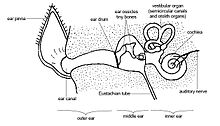
-The rest of the outer ear has bent canals that lead to the eardrum or tympanic membrane. The middle ear is filled with three bones called ossicles and is separated by the outer eardrum in the back of the rabbit's skull.The three ossicles are called hammer, anvil and stirrup and act as a barrier to the inner ear for sound energy, with muscles in the middle ear acting to decrease sound before it hits the inner ear. [1]
- ^ Parsons, Paige K. (2018). "Rabbit Ears: A Structural Look: ...injury or disease, can send your rabbit into a spin". House Rabbit Society.
Week 10: Revised draft[edit]
Introduction to the ears:

The order lagomorphs contain species with adaptions to their anatomical structures whose functions work to help detect and avoid predators. In black tail jack rabbits their long ears cover a great surface area relative to their body size that allows them to detect predators from farther away. In the cotton tailed rabbit, their ears are smaller and shorter which make them have predators be closer to detect them before they flee. In the family leporidae the ears are typically longer than they are wide. Evolution has favored larger rabbits to have shorter ears so the larger surface area does not cause them to lose heat in more temperate regions. The opposite can be seen in rabbits that live in hotter climates, namely because they possess longer ears that help with larger surface area dispersing heat as well as due to the theory that sound does not travel as well in more arid air, opposed to cooler air. Therefore longer ears are meant to aid the organism in detecting prey sooner rather than later in warmer temperatures.[1] The rabbit is characterized by its shorter ears and hindlimbs while hares are characterized by longer ears and longer hindlimbs. [2] Rabbits ears are an important structure to aid in not only in thermoregulation, but also in detecting predators due to how the outer, middle, and inner ear muscles coordinate with one another. The ear muscles also aid in maintaining balance and movement when fleeing predators.

Outer ear (muscles):
The Auricle (anatomy), also known as the pinna is a rabbit's outer ear.[3] The rabbit's body surface is mainly taken up by the pinnae. Their ears, utilized for thermoregulation (particularly the pinnae) contain a vascular network and arteriovenous shunts. It is theorized that the ears aid in losing heat at temperatures above 30°C, with rabbits in warmer climates having longer pinnae due to this. Another theory is that the ears function as shock absorbers that could aid in their vision when running away from predators, but this case has typically only been seen in hares.[4] The rest of the outer ear has bent canals that lead to the eardrum or tympanic membrane.[5]
Middle ear (muscles):
The middle ear is filled with three bones called ossicles and is separated by the outer eardrum in the back of the rabbit's skull.The three ossicles are called hammer, anvil, and stirrup and act as a barrier to the inner ear for sound energy, with muscles in the middle ear acting to decrease sound before it hits the inner ear.[5]
Inner ear (muscles):
Inner ear fluid called endolymph receives the sound energy. Later, within the inner ear there are two parts: cochlea, which utilizes sound waves from the ossicles. and the vestibular apparatus which manages the rabbit's position in regards to movement. Within the cochlea there is a basilar membrane that contains sensory hair structures utilized to send nerve signals to the brain so it can recognize different sound frequencies. Within the vestibular apparatus the rabbit possesses three semicircular canals to help detect angular motion.[5]
***my thermoregulation and pinnae section is meant to go with [[User:Bucl003]] section.
- ^ Hall, E. Raymond. (2001). The Mammals of North America. The Blackburn Press. ISBN 978-1930665354.
- ^ Bensley, Benjamin Arthur (1910). Practical anatomy of the rabbit. The University Press.
- ^ Cite error: The named reference
:3was invoked but never defined (see the help page). - ^ Cite error: The named reference
:4was invoked but never defined (see the help page). - ^ a b c Cite error: The named reference
:5was invoked but never defined (see the help page).
Week 11: Adding an image and caption[edit]

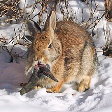
I have selected three different images to utilize for my draft where the first two pictures would be utilized in the introduction to show the physical differences between a hare and a rabbit. Images from our dissection animal were not utilized because the organism is a domestic rabbit and does not rely on their ears as much for fleeing predators or thermoregulation. The third photo shows the muscles interacting in the mammalian ear in general to help the read visualize where all the muscles lie. I could not find any free license or public domain images of muscles in a rabbit's ear therefore the image of the hare was utilized from the scrub hare wiki page, the image of the rabbit was utilized from the Rabbit shopes papilloma virus.jpg wiki image provided in stock, and the anatomy of the mammalian ear was utilized from the anatomy and physiology of animals The ear.jpg wiki image.

Week 12: Going live on Rabbit wiki page[edit]

I added the sentences and heading "thermoregulation" to the rabbit's wikipedia page: "The rabbit's body surface is mainly taken up by the pinnae. Their ears, utilized for thermoregulation (particularly the pinnae) contain a vascular network and arteriovenous shunts[1]" in collaboration with [[User:Bucl003]].
Week 13: Improving/my revising draft[edit]
Introduction to the ears:
The order lagomorphs contain species whose anatomical structures work to help detect and avoid predators. For example, in black tail jack rabbits, their long ears cover a greater surface area relative to their body size that allow them to detect predators from far away. Contrasted to cotton tailed rabbits, their ears are smaller and shorter which force them to have predators be closer to them to be able to detect them before fleeing. In the family leporidae, the ears are typically longer than they are wide. Evolution has favored larger rabbits to have shorter ears so the larger surface area does not cause them to lose heat in more temperate regions. The opposite can be seen in rabbits that live in hotter climates, namely because they possess longer ears that have larger surface area that help with dispersing heat as well as the theory that sound does not travel well in more arid air, opposed to cooler air. Therefore, longer ears are meant to aid the organism in detecting prey sooner rather than later in warmer temperatures.[1] The rabbit is characterized by its shorter ears and hindlimbs while hares are characterized by longer ears and longer hindlimbs. [2] Rabbits ears are an important structure to aid in not only in thermoregulation, but also in detecting predators due to how the outer, middle, and inner ear muscles coordinate with one another. The ear muscles also aid in maintaining balance and movement when fleeing predators.[3]

Outer ear (muscles):
The Auricle (anatomy), also known as the pinna is a rabbit's outer ear.[4] The rabbit's body surface is mainly taken up by the pinnae. Their ears, utilized for thermoregulation (particularly the pinnae) contain a vascular network and arteriovenous shunts. It is theorized that the ears aid in losing heat at temperatures above 30°C, with rabbits in warmer climates having longer pinnae due to this. Another theory is that the ears function as shock absorbers that could aid in their vision when running away from predators, but this case has typically only been seen in hares.[5] The rest of the outer ear has bent canals that lead to the eardrum or tympanic membrane.[6]
Middle ear (muscles):
The middle ear is filled with three bones called ossicles and is separated by the outer eardrum in the back of the rabbit's skull.The three ossicles are called hammer, anvil, and stirrup and act as a barrier to the inner ear for sound energy, with muscles in the middle ear acting to decrease sound before it hits the inner ear.[6]
Inner ear (muscles):
Inner ear fluid called endolymph receives the sound energy. Later, within the inner ear there are two parts: cochlea, which utilizes sound waves from the ossicles. and the vestibular apparatus which manages the rabbit's position in regards to movement. Within the cochlea there is a basilar membrane that contains sensory hair structures utilized to send nerve signals to the brain so it can recognize different sound frequencies. Within the vestibular apparatus the rabbit possesses three semicircular canals to help detect angular motion.[6]
**The next move I will make (in addition to forewarning the Rabbit's talk page), I will coordinate with User:Bucl003 to add my sections of the ears relating to her thermoregulation section. I will then incorporate my introduction to the ears between hares and rabbits in the rabbit's morphology section, but I will post on the talk page about editing the paragraph so our content is not redundant. I will then add my outer, middle, and inner ear sections as well as my mammalian anatomy ear image under an added subsection under Morphology labeled "Anatomy of Ears." In this draft, I have edited grammar and syntax, added a new citation (12), and deleted the images of the hare and rabbit because the Rabbit page already has anatomical structures of the two.
- ^ Cite error: The named reference
:6was invoked but never defined (see the help page). - ^ Cite error: The named reference
:7was invoked but never defined (see the help page). - ^ Meyer, D. L. (1971). "Single Unit Responses of Rabbit Ear-Muscles to Postural and Accelerative Stimulation". Experimental Brain Research. 14 (2): 118–126. doi:10.1007/BF00234795. PMID 5016586. S2CID 6466476.
- ^ Cite error: The named reference
:3was invoked but never defined (see the help page). - ^ Cite error: The named reference
:4was invoked but never defined (see the help page). - ^ a b c Cite error: The named reference
:5was invoked but never defined (see the help page).
Week 14: Final article for rabbit wiki page[edit]
The section I added to the Rabbit wiki page:
Ears[edit]
Introduction to the ears
Within the order lagomorphs, the ears are utilized to detect and avoid predators. In the family leporidae, the ears are typically longer than they are wide. For example, in black tailed jack rabbits, their long ears cover a greater surface area relative to their body size that allow them to detect predators from far away. Contrasted to cotton tailed rabbits, their ears are smaller and shorter, requiring predators to be closer to detect them before fleeing. Evolution has favored rabbits to have shorter ears so the larger surface area does not cause them to lose heat in more temperate regions. The opposite can be seen in rabbits that live in hotter climates, mainly because they possess longer ears that have a larger surface area that help with dispersion of heat as well as the theory that sound does not travel well in more arid air, opposed to cooler air. Therefore, longer ears are meant to aid the organism in detecting prey sooner rather than later in warmer temperatures.[1] The rabbit is characterized by its shorter ears while hares are characterized by their longer ears.[2] Rabbits ears are an important structure to aid thermoregulation and detect predators due to how the outer, middle, and inner ear muscles coordinate with one another. The ear muscles also aid in maintaining balance and movement when fleeing predators.[3]

Outer ear
The Auricle (anatomy), also known as the pinna is a rabbit's outer ear.[4] The rabbit's body surface is mainly taken up by the pinnae. It is theorized that the ears aid in dispersion of heat at temperatures above 30°C with rabbits in warmer climates having longer pinnae due to this. Another theory is that the ears function as shock absorbers that could aid and stabilize rabbit's vision when fleeing predators, but this has typically only been seen in hares.[5] The rest of the outer ear has bent canals that lead to the eardrum or tympanic membrane.[6]
Middle ear
The middle ear is filled with three bones called ossicles and is separated by the outer eardrum in the back of the rabbit's skull.The three ossicles are called hammer, anvil, and stirrup and act to decrease sound before it hits the inner ear. In general, the ossicles act as a barrier to the inner ear for sound energy. [6]
Inner ear
Inner ear fluid called endolymph receives the sound energy. After receiving the energy, later within the inner ear there are two parts: the cochlea that utilizes sound waves from the ossicles and the vestibular apparatus that manages the rabbit's position in regards to movement. Within the cochlea there is a basilar membrane that contains sensory hair structures utilized to send nerve signals to the brain so it can recognize different sound frequencies. Within the vestibular apparatus the rabbit possesses three semicircular canals to help detect angular motion.[6]
*I also added this sentence to [[User:Bucl003]] Thermoregulation section: This process is carried out by the pinnae which takes up most of the rabbit's body surface and contain a vascular network and arteriovenous shunts.[7]
- ^ Hall, E. Raymond. (2001). The Mammals of North America. The Blackburn Press. ISBN 978-1930665354.
- ^ Bensley, Benjamin Arthur (1910). Practical anatomy of the rabbit. The University Press.
- ^ Meyer, D. L. (1971). "Single Unit Responses of Rabbit Ear-Muscles to Postural and Accelerative Stimulation". Experimental Brain Research. 14 (2): 118–126. doi:10.1007/BF00234795. PMID 5016586. S2CID 6466476.
- ^ Capello, Vittorio (2006). "Lateral Ear Canal Resection and Ablation in Pet Rabbits" (PDF). The North American Veterinary Conference. 20.
- ^ Vella, David (2012). Ferrets, Rabbits, and Rodents: Clinical Medicine and Surgery. Elsevier. ISBN 978-1-4160-6621-7.
- ^ a b c Parsons, Paige K. (2018). "Rabbit Ears: A Structural Look: ...injury or disease, can send your rabbit into a spin". House Rabbit Society.
- ^ Vella, David (2012). Ferrets, Rabbits, and Rodents: Clinical, Medicine, and Surgery. Elsevier. ISBN 9781416066217.
