Wikipedia:Featured picture candidates/Human Blood Cells
Human Blood Cells[edit]

- Reason
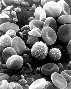
- The image is high resolution, depicting in good detail the cells found in human blood, adding much to the topics using it
- Articles this image appears in
- Immune system, Scanning electron microscope, White blood cell, Complete blood count, Lymphocyte, Monocyte, Innate immune system
- Creator
- User:DO11.10
- Support as nominator --J6kyll (talk) 03:37, 25 October 2008 (UTC)
- oppose much more detailed images of cells are possible. [1]. de Bivort 04:30, 25 October 2008 (UTC)
- Oppose - far too unsharp for what an SEM is capable of. To note, it was created in 1982 - technology in this area has improved since then. —Vanderdecken∴ ∫ξφ 08:51, 25 October 2008 (UTC)
- Oppose and speedy close as per WP:SNOW. The image is lacking in sharpness: the detail present just isn't enough. I say speedy close. Elucidate (parlez à moi) Ici pour humor 20:52, 25 October 2008 (UTC)
- I don't think three opposes is enough for a speedy close tbh. The nominator might get scared that we're speedily removing it as if we think it's obvious to anyone that it's not up to standard and it's not worth our time - they might not have realised how much better than this SEM pics can be. —Vanderdecken∴ ∫ξφ 09:10, 26 October 2008 (UTC)
- Oppose I agree with Vanderdecken, SEM pics can be much better. SpencerT♦C 21:11, 28 October 2008 (UTC)
Not promoted --Elucidate (parlez à moi) Ici pour humor 12:26, 30 October 2008 (UTC)
