User:Jyangmidd/sandbox
This is my sand box.
I am practicing writing for Introduction to Neuroscience at Middlebury College. Middlebury College
My Contribution to the Inferior Temporal Cortex Page[edit]
Receiving Information[edit]
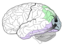
The light energy that comes from the rays bouncing off of an object is converted into chemical energy by the cells in the retina of the eye. This chemical energy is then converted into action potentials that are transferred through the optic nerve and across the optic chiasm, where it is first processed by the lateral geniculate nucleus of the thalamus. From there the information is sent to the primary visual cortex, region V1. It then travels from the visual areas in the occipital lobe to the parietal and temporal lobes via two distinct anatomical streams. [1] These two cortical visual systems were classified by Ungerleider and Mishkin (1982, see two-streams hypothesis)[2] . One stream travels ventrally to the inferior temporal cortex (from V1 to V2 then through V4 to ITC) while the other travels dorsally to the posterior parietal cortex. They are labeled the “what” and “where” streams , respectively. The Inferior Temporal Cortex receives information from the ventral stream, understandably so, as it is known to be a region essential in recognizing patterns, faces, and objects.[3]

How it Works[edit]
The information for color and form comes from P-cells that receive their information mainly from cones, so they are sensitive to differences in form and color, as opposed to the M-cells that receive information about motion mainly from rods. The neurons in the inferior temporal cortex, also called the inferior temporal visual association cortex, process this information from the P-cells. [4] The neurons in the ITC have several unique properties that offer an explanation as to why this area is essential in recognizing patterns. They only respond to visual stimuli and their receptive fields always include the fovea, which is one of the densest areas of the retina and is responsible for acute central vision. These receptive fields tend to be larger than those in the striate cortex and often extend across the midline to unite the two visual half fields for the first time. IT neurons are selective for shape and/or color of stimulus and are usually more responsive to complex shapes as opposed to simple ones. A small percentage of them are selective for specific parts of the face. Faces and likely other complex shapes are seemingly coded by a sequence of activity across a group of cells, and IT cells can display both short or long term memory for visual stimuli based on experience. [5]
Object Recognition[edit]
There are a number of regions within the ITC that work together for processing and recognizing the information of “what” something is. In fact, discrete categories of objects are even associated with different regions.
- The fusiform gyrus or Fusiform Face Area (FFA) deals more with facial and body recognition rather than objects.

- The Parahippocampal Place Area (PPA) helps differentiate between scenes and objects.

- The Extrastriate Body Area (EBA) tells apart body parts from other objects
- And the Lateral Occipital Complex (LOC) is used to determine shapes vs. scrambled stimuli.
These areas must all work together, as well as with the hippocampus, in order to create an array of understanding of the physical world. The hippocampus is key for storing the memory of what an object is/what it looks like for future use so that it can be compared and contrasted with other objects. Correctly being able to recognize an object is highly dependent on this organized network of brain areas that process, share, and store information. In a study by Denys et al., functional magnetic resonance imaging (FMRI) was used to compare the processing of visual shape between humans and macaques. They found, amongst other things, that there was a degree of overlap between shape and motion sensitive regions of the cortex, but that the overlap was more distinct in humans. This would suggest that the human brain is better evolved for a high level of functioning in a distinct, three-dimensional, visual world.[7]
See Also[edit]
Cognitive Neuroscience of Visual Object Recognition
Neural Processing for Individual Categories of Objects
References[edit]
- ^ Kolb, Bryan; Whishaw, Ian Q. (2014). An Introduction to Brain and Behavior (Fourth ed.). New York, NY: Worth. pp. 282–312.
{{cite book}}: CS1 maint: multiple names: authors list (link) - ^ Mishkin, Mortimer; Ungerleider, Leslie G. (1982). "Two Cortical Visual Systems" (PDF). The MIT Press.
{{cite journal}}: Cite journal requires|journal=(help)CS1 maint: multiple names: authors list (link) - ^ Creem, Sarah H.; Proffitt, Dennis R. (2001). "Defining the cortical visual systems: "What", "Where", and "How"" (PDF). Acta Psychologica (107): 43–68.
{{cite journal}}: CS1 maint: multiple names: authors list (link) - ^ Dragoi, Valentin. "Chapter 15: Visual Processing: Cortical Pathways". Retrieved 12 November 2013.
- ^ Gross, Charles. "Inferior temporal cortex". Retrieved 12 November 2013.
- ^ Spiridon, M., Fischl, B., & Kanwisher, N. (2006). Location and spatial profile of category-specific regions in human extrastriate cortex. Human Brain Mapping , 27, 77-89.
- ^ Denys; et al. (March 10, 2004). "The Processing of Visual Shape in the Cerebral Cortex of Human and Nonhuman Primates: A Functional Magnetic Resonance Imaging Study" (PDF). The Journal of Neuroscience: 2551–2565.
{{cite journal}}: Explicit use of et al. in:|last=(help)
