
File:Axial twist in zebrafish embryo.pdf

Size of this JPG preview of this PDF file: 800 × 575 pixels. Other resolutions: 320 × 230 pixels | 640 × 460 pixels | 1,024 × 737 pixels | 1,204 × 866 pixels.
Original file (1,204 × 866 pixels, file size: 33 KB, MIME type: application/pdf)
| This is a file from the Wikimedia Commons. Information from its description page there is shown below. Commons is a freely licensed media file repository. You can help. |
Summary
| Description |
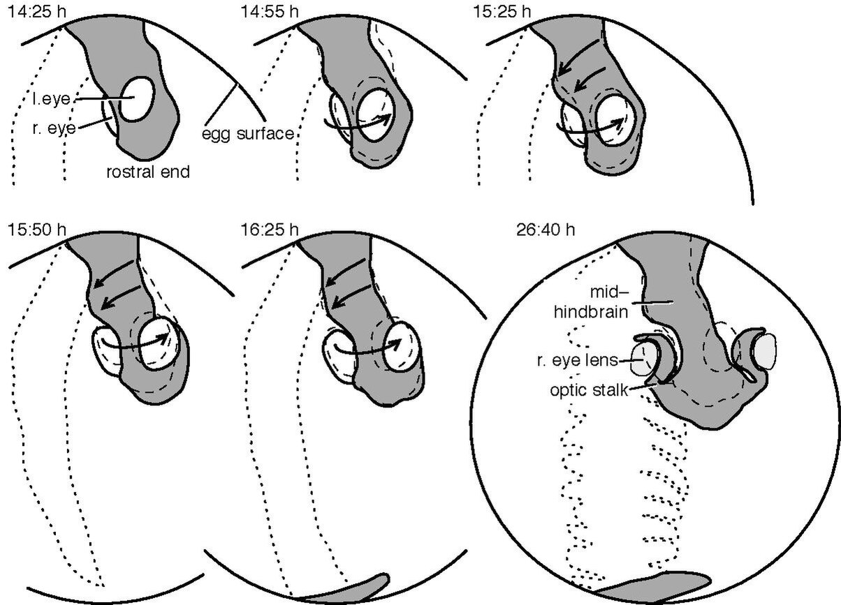
English: The embryo is drawn in grey, the prospective eye regions white. Dashed contours show the previous location of the embryo. The location of the body on the back side of the egg is drawn dotted. Compensatory movements can be observed between 14:40 and 16:40 p.f. During this period those cells that will form the eyes migrate anti-clockwise (perspective of the embryo), whereas the future mid- and hindbrain cells migrate clockwise between 15:15 and 16:40 h (arrows). The right eye is initially invisible because it is hidden below the cells that will form the forebrain. The first 5 frames are interleaved by 30 min, the last one is 10:15 h later. Drawn from Keller et al. (2008: supplementary movie no. 2).
See also: https://figshare.com/articles/media/An_embryological_twist_in_zebra_fish_embryo_as_revealed_by_cellular_movement_patterns_/24104085?file=42290463 This is Figure 4 from: Marc H. E. de Lussanet and Jan W. M. Osse. An ancestral axial twist explains the contralateral forebrain and the optic chiasm in vertebrates. Animal Biol., 62(2):193–216, 2012. doi: 10.1163/157075611X617102. URL http://arxiv.org/abs/1003.1872. The publisher, BRILL, Leiden, Netherlands, kindly granted sharing the figure on Wikipedia. |
| Date | |
| Source | Own work |
| Author | Marc HE de Lussanet |
Licensing
I, the copyright holder of this work, hereby publish it under the following license:
| This file is made available under the Creative Commons CC0 1.0 Universal Public Domain Dedication. | |
| The person who associated a work with this deed has dedicated the work to the public domain by waiving all of their rights to the work worldwide under copyright law, including all related and neighboring rights, to the extent allowed by law. You can copy, modify, distribute and perform the work, even for commercial purposes, all without asking permission.
|
File history
Click on a date/time to view the file as it appeared at that time.
| Date/Time | Thumbnail | Dimensions | User | Comment | |
|---|---|---|---|---|---|
| current | 20:05, 9 September 2023 |  | 1,204 × 866 (33 KB) | Marci68 | Uploaded own work with UploadWizard |
File usage
The following pages on the English Wikipedia use this file (pages on other projects are not listed):