
Supratrochlear nerve
| Supratrochlear nerve | |
|---|---|
 Sensory areas of the head, showing the general distribution of the three divisions of the fifth nerve. (Supratrochlear nerve labeled at upper left.) | |
 Nerves of the orbit. Seen from above. (Supratrochlear nerve visible near top.) | |
| Details | |
| From | Frontal nerve |
| Identifiers | |
| Latin | nervus supratrochlearis |
| TA98 | A14.2.01.024 |
| TA2 | 6203 |
| FMA | 52642 |
| Anatomical terms of neuroanatomy | |
The supratrochlear nerve is a branch of the frontal nerve, itself a branch of the ophthalmic nerve (CN V1) from the trigeminal nerve (CN V). It provides sensory innervation to the skin of the forehead and the upper eyelid.
Structure[edit]
Origin[edit]
The supratrochlear nerve is the smaller of the two terminal branches of the frontal nerve (the other being the supraorbital nerve).[1] It arises midway between the base and apex of the orbit[2] where the frontal nerve splits into said terminal branches.[1]
Course[edit]
The supratrochlear nerve passes medially[3] above the trochlea of the superior oblique muscle.[2][3] It then travels anteriorly above the levator palpebrae superioris muscle.[1] It exits the orbit through the supraorbital notch or foramen.[3] It then ascends onto the forehead beneath the corrugator supercilii muscle and frontalis muscle. It finally divides into sensory branches.[citation needed]
The supratrochlear nerve travels with the supratrochlear artery, a branch of the ophthalmic artery.[2]
Branches[edit]
Before exiting the orbit, the supratrochlear nerve emits a descending branch to the infratrochlear nerve.[3][1]
Function[edit]
The supratrochlear nerve provides sensory innervation to the skin and conjunctiva of the upper eyelid, and the skin of the inferomedial forehead.[1] It may also provide sensory innervation to part of the periosteum of the frontal bone.[4]
Clinical significance[edit]
The supratrochlear nerve may be anaesthetised for surgery of parts of the scalp.[5][6] This can be used for small lesions of the scalp.[5] It can also be used for more extensive injury to the scalp.[6] It is often anaesthetised alongside the supraorbital artery.[5]
Etymology[edit]
The supratrochlear nerve is named for its passage above the trochlea of the superior oblique muscle.[2]
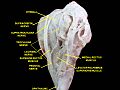
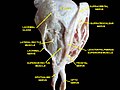
Additional images[edit]
-
 Supratrochlear nerve
 -
 Extrinsic eye muscle. Nerves of orbita. Deep dissection.
 -
 Extrinsic eye muscle. Nerves of orbita. Deep dissection.
 -
 Extrinsic eye muscle. Nerves of orbita. Deep dissection.
 -
 Extrinsic eye muscle. Nerves of orbita. Deep dissection.
 -
 Extrinsic eye muscle. Nerves of orbita. Deep dissection.


References[edit]
![]() This article incorporates text in the public domain from page 888 of the 20th edition of Gray's Anatomy (1918)
This article incorporates text in the public domain from page 888 of the 20th edition of Gray's Anatomy (1918)
- ^ a b c d e Fillmore, Erin P.; Seifert, Mark F. (2015). "22 - Anatomy of the Trigeminal Nerve". Nerves and Nerve Injuries. Vol. 1: History, Embryology, Anatomy, Imaging, and Diagnostics. Academic Press. pp. 319–350. doi:10.1016/B978-0-12-410390-0.00023-8. ISBN 978-0-12-410390-0.
- ^ a b c d Rea, Paul (2016). "2 - Head". Essential Clinically Applied Anatomy of the Peripheral Nervous System in the Head and Neck. Academic Press. pp. 21–130. doi:10.1016/B978-0-12-803633-4.00002-8. ISBN 978-0-12-803633-4.
- ^ a b c d Standring, Susan (2020). Gray's Anatomy: The Anatomical Basis of Clinical Practice (42nd ed.). New York. p. 782. ISBN 978-0-7020-7707-4. OCLC 1201341621.
{{cite book}}: CS1 maint: location missing publisher (link) - ^ Barral, Jean-Pierre; Croibier, Alain (2009). "15 - Ophthalmic nerve". Manual Therapy for the Cranial Nerves. Churchill Livingstone. pp. 115–128. doi:10.1016/B978-0-7020-3100-7.50018-5. ISBN 978-0-7020-3100-7.
- ^ a b c Kinder Ross, Alison; Bryskin, Robert B. (2011). "16 - Regional Anesthesia". Smith's Anesthesia for Infants and Children (8th ed.). Mosby (imprint). pp. 452–510. doi:10.1016/B978-0-323-06612-9.00016-X. ISBN 978-0-323-06612-9.
- ^ a b Trott, Alexander T. (2012). "6 - Infiltration and Nerve Block Anesthesia". Wounds and Lacerations - Emergency Care and Closure (4th ed.). Saunders. pp. 41–72. doi:10.1016/B978-0-323-07418-6.00006-X. ISBN 978-0-323-07418-6.
External links[edit]
- Anatomy figure: 29:02-01 at Human Anatomy Online, SUNY Downstate Medical Center
- MedEd at Loyola GrossAnatomy/h_n/cn/cn1/cnb1.htm
- lesson3 at The Anatomy Lesson by Wesley Norman (Georgetown University) (orbit2)
- cranialnerves at The Anatomy Lesson by Wesley Norman (Georgetown University) (V)
- http://www.dartmouth.edu/~humananatomy/figures/chapter_47/47-2.HTM