
File:Rubella virus 10221 lores.jpg

Size of this preview: 447 × 599 pixels. Other resolutions: 179 × 240 pixels | 358 × 480 pixels | 573 × 768 pixels | 764 × 1,024 pixels | 2,341 × 3,136 pixels.
Original file (2,341 × 3,136 pixels, file size: 2.01 MB, MIME type: image/jpeg)
| This is a file from the Wikimedia Commons. Information from its description page there is shown below. Commons is a freely licensed media file repository. You can help. |
Summary
| Description |
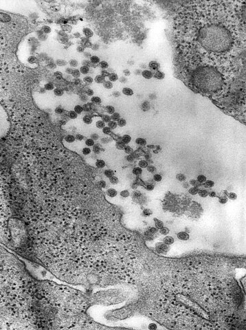
English: This negatively-stained transmission electron micrograph (TEM) revealed the presence of Rubella virus virions, as they were in the process of budding from the host cell surface to be freed into the host’s system, thereby, producing an enveloped virus particle, which means that after budding, the spherical virions' icosahedral capsid is enclosed in the host cell membrane. Inside the capsid lies the Rubella virus’ positive-sense single-stranded RNA ((+)ssRNA) genome. The Rubella virus is known to be the cause of rubella, otherwise known as German measles.
Deutsch: Transmissions-Elektronenmikroskopische (TEM) Aufnahme von Rubella-Viren, die gerade von der Zelloberfläche ausknospen und in den Wirtsorgan freigesetzt werden. Dabei entsteht ein behüllter Virus-Partikel, bei dem das ikosaedrische Kapsid von der Zellmembran der Wirtszelle umhüllt ist. Im Inneren des Kapsids liegt die Einzelstrang-RNA des Virus. Rubella-Virus ist der Erreger der Röteln. |
|||
| Date | ||||
| Source | http://phil.cdc.gov/phil/home.asp ID# 10221 US Department of Health and Human Services | |||
| Author | CDC/ Dr. Fred Murphy; Sylvia Whitfield | |||
| Permission (Reusing this file) |
|
Licensing
|
This media comes from the Centers for Disease Control and Prevention's Public Health Image Library (PHIL), with identification number #10221. Note: Not all PHIL images are public domain; be sure to check copyright status and credit authors and content providers.
|
File history
Click on a date/time to view the file as it appeared at that time.
| Date/Time | Thumbnail | Dimensions | User | Comment | |
|---|---|---|---|---|---|
| current | 20:56, 2 November 2016 | | 2,341 × 3,136 (2.01 MB) | Opencooper | higher resolution |
| 08:50, 22 October 2008 |  | 700 × 937 (145 KB) | Der Lange | {{Information |Description={{en|1=This negatively-stained transmission electron micrograph (TEM) revealed the presence of Rubella virus virions, as they were in the process of budding from the host cell surface to be freed into the host’s system, thereb |
File usage
The following pages on the English Wikipedia use this file (pages on other projects are not listed):
Global file usage
The following other wikis use this file:
- Usage on en.wiktionary.org
- Usage on jv.wikipedia.org
- Usage on ml.wikipedia.org