
File:3D ribbon diagram of alpha-neurexin 1.png
No higher resolution available.
3D_ribbon_diagram_of_alpha-neurexin_1.png (357 × 279 pixels, file size: 60 KB, MIME type: image/png)
Summary[edit]
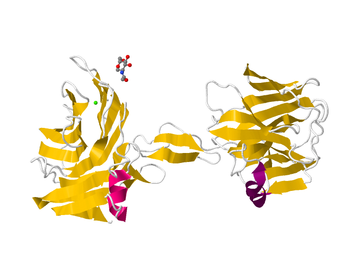
| Description | This is a 3D ribbon diagram of an alpha-neurexin protein. It shows the secondary structure and ligand. |
|---|---|
| Author or copyright owner |
Protein Databank |
| Source (WP:NFCC#4) | http://www.rcsb.org/pdb/explore/jmol.do?structureId=3ASI&bionumber=1 |
| Use in article (WP:NFCC#7) | Neurexin |
| Purpose of use in article (WP:NFCC#8) | It gives a visual image of the protein discussed in the article. One can grasp more fully how this protein works if one can see what it looks like. |
| Not replaceable with free media because (WP:NFCC#1) |
n.a. |
| Minimal use (WP:NFCC#3) | It will be used once on this page. |
| Respect for commercial opportunities (WP:NFCC#2) |
n.a. |
| Fair useFair use of copyrighted material in the context of Neurexin//en.wikipedia.org/wiki/File:3D_ribbon_diagram_of_alpha-neurexin_1.pngtrue | |
Licensing[edit]
 | This is a two-dimensional representation of a copyrighted sculpture, statue or any other three-dimensional work of art. As such it is a derivative work of art, and per US Copyright Act of 1976, § 106(2) whoever holds copyright of the original has the exclusive right to authorize derivative works. Per § 107 it is believed that reproduction for criticism, comment, teaching and scholarship constitutes fair use and does not infringe copyright. It is believed that the use of a picture
qualifies as fair use under the Copyright law of the United States. Any other uses of this image, on Wikipedia or elsewhere, might be copyright infringement. |
File history
Click on a date/time to view the file as it appeared at that time.
| Date/Time | Thumbnail | Dimensions | User | Comment | |
|---|---|---|---|---|---|
| current | 19:23, 22 February 2017 | | 357 × 279 (60 KB) | DatBot (talk | contribs) | Reduce size of non-free image (BOT - disable) |
| 19:56, 12 April 2013 | No thumbnail | 752 × 589 (153 KB) | Rachelbash1 (talk | contribs) | Uploading an excerpt from a non-free work using File Upload Wizard |
You cannot overwrite this file.
File usage
The following pages on the English Wikipedia use this file (pages on other projects are not listed):